Talk to an Expert
Talk to an Expert
If you're looking for something, we can help! Give us a call at 1 (888) 228-7564 or shoot us an email anytime: Sales@IntegrisEquipment.com
Extended Main Unit warranty for 1 additional year of coverage for SoloScan. Up to 3 additional years may be purchased (for main unit only)
Linear probe for vascular, small parts, breast, MSK, nerve, anesthesia
Small Linear probe for nerve, anesthesia, vascular, small parts, breast, MSK
Transvaginal probe style for obstetrics/Gynecology, Urology, IVF, Pelvic
Micro-Convex probe for neonatal, abdominal, small parts, neonatal cardiac 6.5 MHZ
Dual Convex/Cardiac probe style fo emergency cardiac abdomen, ob/gyn, urology, lung
Convex probe for abdomen, ob/gyn, urology, lung
P50 Handheld Ultrasound. Main unit only- to be used with interchangable probe heads. WiFi and USB connection supported by iOS, Android, and Windows...
View full detailsNeedle Guide Bracket for Micro-convex array transducer C6152UB, BGK-R15UB.
Needle Guide Bracket for linear transducer L742UB/L1042UB, BGK-L40UB.
Needle Guide Bracket for Linear array transducer L552UB, BGK-L50UB.
Needle Guide Bracket for Micro-convex array transducer: C422UB, BGK-R20UB.
Needle Guide Bracket for Micro-convex array transducer C612UB , BGK-R10UB.
Needle Guide Bracket for transvaginal transducer E611-2/E8-4Q/E8-4D, BGK-CR10UA
Needle Guide Bracket for convex transducer C5-2b/C5-2XQ/C5-2Q/C5-2XD/C5-2D, BGK-C5-2
Needle Guide Bracket for convex transducer C5-2b, BGK-C5-2.
Needle Guide Bracket for convex transducer C352UB, BGK-R50UB.
Needle Guide Bracket for micro-convex transducer C613UA, BGK-MCR10.
Needle Guide Bracket for linear transducer L763UA, BGK-LA70.
Needle Guide Bracket for transrectal transducer E743UA, BGK-EL40.
Needle Guide Bracket for Transvaginal transducer E613UA, BGK-CR10UA.
Needle Guide Bracket for micro-convex transducer C321UA, BGK-CR20.
Needle Guide Bracket for linear transducer L743UA&L742UA, BGK-LA43.
Needle Guide Bracket for convex transducer C343UA, BGK-CR40.
What is Ultrasound?
Ultrasound is cyclic sound pressure with a frequency greater than the upper limit of human hearing. Ultrasound is thus not separated from "normal" (audible) sound based on differences in physical properties, only the fact that humans cannot hear it. Although this limit varies from person to person, it is approximately 20 kilohertz (20,000 hertz) in healthy, young adults. The production of ultrasound is used in many different fields, typically to penetrate a medium and measure the reflection signature or supply focused energy. The reflection signature can reveal details about the inner structure of the medium, a property also used by animals such as bats for hunting. The most well known application of ultrasound is its use in sonography to produce pictures of fetuses in the human womb. There are a vast number of other applications as well.
Medical sonography (ultrasonography) is an ultrasound-based diagnostic medical imaging technique used to visualize muscles, tendons, and many internal organs, to capture their size, structure and any pathological lesions with real time tomographic images. Ultrasound has been used by radiologists and sonographers to image the human body for at least 50 years and has become one of the most widely used diagnostic tools in modern medicine. The technology is relatively inexpensive and portable, especially when compared with other techniques, such as magnetic resonance imaging (MRI) and computed tomography (CT). Ultrasound is also used to visualize fetuses during routine and emergency prenatal care. Such diagnostic applications used during pregnancy are referred to as obstetric sonography.
Obstetric ultrasound can be used to identify many conditions that would be harmful to the mother and the baby. Many health care professionals consider the risk of leaving these conditions undiagnosed to be much greater than the very small risk, if any, associated with undergoing an ultrasound scan. Sonography is used routinely in obstetric appointments during pregnancy, but the FDA discourages its use for non-medical purposes such as fetal keepsake videos and photos, even though it is the same technology used in hospitals.
Obstetric ultrasound is primarily used to:
Ultrasound scanners have different Doppler-techniques to visualize arteries and veins. The most common is colour doppler or power doppler, but also other techniques like b-flow are used to show bloodflow in an organ. By using pulsed wave doppler or continuous wave doppler bloodflow velocities can be calculated.
Ultrasound is also increasingly being used in trauma and first aid cases, with emergency ultrasound becoming a staple of most EMT response teams. Furthermore, ultrasound is used in remote diagnosis cases where teleconsultation is required, such as scientific experiments in space or mobile sports team diagnosis.
For any Ultrasound Equipment not listed here, please do not hesitate to call or email!!
888-228-7564
Ultraclean Elec:1"Ndle;Ext In Free Shipping
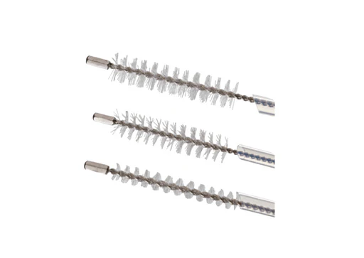
Duodeno 3mm Brush Sheath Diameter - 1.80mm Sheath Length - 200.00cm Purchasing Unit of Measure - Qty 20 Free Shipping
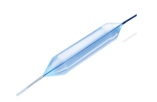
18mm X 8CM Esoph Bal DilCONMED's Eliminator® PET Balloon Dilators offer a simple solution capable of dilating the tightest of strictures. Construct...
View full detailsVyaire Medical Airlife® Disposable Self-Inflating Resuscitation Devices - Vyaire CRF-2K8018Resuscitation Device, Pediatric, w/ Mask, 28" Large Bore...
View full detailsClear recently viewed items
Reviews
Arrived very quickly. It was just what I was looking for and with a more approachable value.
Great timely service, will use again.
Great replacement battery!, it even has an expiration on it so that I know when to change it in 5 years. Last ones I purchased from another seller was garbage.
Good turn around time
Great product and service
Everything is as expected.


